Author: Craig Steele

Three Reasons to see a Physiotherapist

Most people associate physiotherapy with pain and injury management.  While helping you recover from pain is our specialty, physiotherapists are also able to help with many more issues. Here are three things that you may not have thought to visit a physiotherapist for.


Stiffness and Inflexibility


Almost all of us have experienced pain and stiffness after a day of increased or unaccustomed exercise. This kind of stiffness usually wears of quickly, and is referred to as DOMS (delayed onset muscles soreness). If however, you find yourself feeling stiff for longer periods, or even most the time – it might be time to see a physiotherapist. There are many different causes of stiffness and inflexibility; by far the most common is lack of movement. Our joints and muscles both lose flexibility if they are not regularly moved all the way through their range. Muscles can feel short and tight with a bouncy feeling of restriction and joints are more of a hard ‘blocked’ feeling when you try to move.


For this kind of stiffness, you may not even notice that you have lost range, as it can be very easy to adapt your movements to compensate. Your physiotherapist can help you to identify where you have areas of inflexibility and help you to exercise, stretch and mobilise your joints to get them back to a healthy range. Disease processes such as Osteoarthritis and Rheumatoid Arthritis can also cause prolonged stiffness and your physiotherapist is well equipped to help you deal with these conditions.


Reduced Strength or Weakness


There are many reasons for weakness in the body, from generalised disuse, weakness in one muscle group following injury, neurological weakness or structural weakness of joint following an injury. Weakness of any kind can predispose you to future injury and can be surprisingly difficult to resolve without targeted exercises. Your physiotherapist is able to determine the cause of your weakness and determine the best treatment to restore your muscle strength.

Reduced Balance

Keeping your balance is a very complicated process and your body works hard to make sure you stay on your feet. Humans have a very small base of support for our height and we use all our senses together to determine which movements we should make to stay upright, including our visual, vestibular, muscular and sensory systems. As balance is so important, if one part of our senses begins to weaken, the others will quickly compensate, so you may not notice that your balance has worsened until you fall or trip over.

As a general rule, our balance deteriorates as we age but this does not mean that falls should be an inevitable part of aging. Actively working to maintain or improve your balance can have a significant effect on your quality of life and confidence in getting around. Your physiotherapist is able to test all the aspects of your balance and provide effective rehabilitation to help keep you on your feet.

Osteoarthritis of the Hip

Osteoarthritis (OA) is a degenerative disease that affects the cartilage of joints. Cartilage is a firm, flexible connective tissue that lines the surface of many joints and provides shock absorption and cushioning for the bony surfaces of those joints as they move. During the process of OA, cartilage gradually begins to break down and is worn away. This means that the bony surfaces below the cartilage start to rub together, creating increased stress and friction. The body reacts to this increased stress by creating small bony deposits around the joint, as more of these are created the joint becomes increasingly painful and difficult to move.

The hip is one the joints most commonly affected by osteoarthritis. While OA is generally considered to be a disease associated with aging, younger people can be affected, particularly following trauma to the hip. As a general rule, however, the cartilage in our bodies loses elasticity as we age, making it more susceptible to damage. Other risk factors for the development of OA are a family history of OA, previous traumatic injury of the hip, obesity, improper formation of the hip at birth (developmental dysplasia), genetic defects of the cartilage, impingement of the hip (femoroacetabular impingement) and a history of intense weight bearing activities.

What are the signs and symptoms?

The most common symptoms of hip OA are pain and stiffness with reduced movement of the hip, particularly in the direction of internal rotation. These symptoms in a person over the age of 50, in the absence of a trauma that may have caused a fracture, indicate possible OA. Pain originating from the hip joint can be felt as a deep ache that can be noticed in the groin, buttocks, thigh or even knee. It is also typical for sufferers of OA to experience stiffness in the morning upon waking that lasts less than 30-60 minutes. Grating or cracking sensations with hip movements are also common complaints, along with mild to moderate joint swelling.

In the early stages, mild pain may be felt with activities such as walking or running. As the disease progresses these activities will become more painful with the muscles that provide additional support to the joint becoming weaker, exacerbating the disease process. For many people, a total hip replacement may be necessary to reduce pain and restore function.

How can physiotherapy help?

For mild to moderate cases of OA, physiotherapy can help to reduce pain and maintain function for as long as possible. Keep the musculature around the hip as Strong and healthy as possible can have a significant impact on your quality of life and your physiotherapist work with you to help you to set and reach your goals for treatment
Treatment will also include stretching, trigger point therapy, joint mobilization to increase the joint’s mobility, and a personalised exercise program, including hydrotherapy and isometric exercises that work to increase muscle strength while putting less pressure on the joint.

For those whose best course of treatment is surgical joint replacement, physiotherapy can help to achieve great outcomes by helping with effective preparation and rehabilitation, getting you on your way to recovery as quickly as possible.

None of the information in this article is a replacement for proper medical advice. Always see a medical professional for advice on your individual condition.

What are spondylolysis and spondylolisthesis

One of the primary roles of the spine is to protect the spinal cord. This means that the spine needs to be strong while maintaining the flexibility required for a movable trunk. While the spine is very sturdy, spinal injuries do occur. Health professionals often use terms to describe and classify injuries of the body, two of these terms that you may have heard are Spondylolysis and Spondylolisthesis.

What are they?

Spondylolysis refers to a stress fracture of the pars interarticularis of the vertebra. This is the part of the vertebra that connects the body of the vertebra with the rest of the vertebra that surrounds the spinal cord. A separation of this fracture where the body of the vertebra is displaced forwards or backwards is called a spondylolisthesis.

Spondylolisthesis is a progression of spondylolysis and is given grades to classify its severity. Both spondylolysis and spondylolisthesis commonly affect the fourth and fifth lumbar vertebrae, found at the base of the lower back.

What are the causes?

Spondylolysis and spondylolisthesis can be a result of trauma with the spine being moved forcefully into extension, particularly in younger people. Certain sports such as gymnastics, football and weightlifting require repetitive backward movements of the spine and this can eventually lead to a stress fracture of the pars interarticularis. Growth spurts in teens have also been known to be responsible for the development of these conditions.


In older adults, common causes of spondylolysis or spondylolisthesis are degenerative changes in the spine due to aging, osteoporosis, infection or even a tumour. Some people have a genetic vulnerability in this area of their spine making them more susceptible to developing spondylolysis and then spondylolisthesis.


What are the symptoms?


Many people with spondylolysis and spondylolisthesis may be asymptomatic, which means they perform their normal activities without experiencing any symptoms. However, when symptoms do occur, common complaints are pain and tightness, much like a muscle strain, spreading across the lower back. This pain may be eased by bending forwards and aggravated by walking, running or leaning backwards.


In more progressive cases of spondylolisthesis, the shift of the vertebral body can cause narrowing of the spinal canal that can lead to nerve compression. This may cause hamstring tightness and even numbness and weakness of the lower limbs, affecting gait and daily activities.


How can physiotherapy help?


Your physiotherapist will work closely with you and any relevant medical professionals to determine exactly what is needed for your particular condition. Severe instability in the spine may require stabilization surgery, however this is rare and in most cases symptoms of spondylolisthesis can be improved with regular physiotherapy management.


Physiotherapy that focuses on strengthening and improving the flexibility of both the lower back and the abdominal muscles has been shown to have positive effects on both pain and function for those with symptomatic spondylolysis and spondylolisthesis.


Speak to your physiotherapists for more information regarding your individual condition. None of the information in this newsletter is a replacement for proper medical advice. Always see a medical professional for advice on your individual injury.

Heel Pain – Plantar Fasciitis

Plantar fasciitis is a common condition causing pain in the heel or into the arch of the foot. The Plantar fascia is a connective tissue which runs underneath the foot from the heel bone to the base of the toes forming the foot’s arch. Often injury is due to repetitive load and less commonly due to a traumatic injury. Due to the insertion of the plantar fascia on the heel bone often it will present as heel pain on walking.
Causes:
Repetitive or prolonged activities i.e. runners, dancers, gymnasts
Sudden increase in exercise or load
Poor foot posture- flat feet or high arches
Poor hip and knee muscle strength and control
Calf muscle tightness or joint stiffness
Inappropriate footwear
Signs and symptoms to be aware of are morning pain with the first few steps which eases with movement, an initial onset of pain with exercise which eases as you warm up, tenderness on the arch of the foot or the heel and pain when walking with no shoes or flat soles. With a progression of symptoms people may experience constant pain with walking which increases with loading and exercise.
A Physiotherapist will be able to diagnose plantar fasciitis by thorough assessment to target the cause and best management approach individually for you. Treatment may consist of advise on appropriate footwear, arch support when required, soft tissue release, joint mobilisation and home exercises of stretching and muscle strengthening. A Physio is then able to rehabilitate a patient through a gradual return to exercise to prevent reoccurrence of the injury.
If you have been experiencing heel or arch pain a physiotherapist at Hinteractive Physio can assess and diagnose the source of your symptoms to assist with the best treatment and management. Call us today on 54425556 or email us hinteractivephyio@gmail.com is you have any questions of enquires.

What is Yin Yoga?

Blog by Physiotherapist Simone Bifulco

Most forms of Yoga today are dynamic and very active in their practice, designed to work more on the muscular part of the body.  Yin Yoga is opposite to this.  Yin Yoga works on another deeper layer of our soft tissues called fascia.  It is a gentle but very powerful practice that works deeper into the fascial network system of our bodies.  Yang is everything that moves and is symbolized by fire and sun.  Yin is the slow, the calm energy, the water, the moon.  Everybody has the Yin and Yang energy Chi within themselves.  The Yin Yoga practice consists of specific poses that are held in stillness for between 3 and 5 minutes to promote an elongation of the fascia being worked on in those poses and activating the energy chi to flow in those areas as well.  Yin Yoga goes beyond a physical workout and helps us to balance out energetically, emotionally and mentally.

Osgood Schlatters – The Sporting Knee

The Sporting knee: Osgood Shlatters
Osgood Shlatters is an overuse injury of the knee which commonly affects children and adolescents. Symptoms usually develop during a growth spurt due to an increase in tension on the tendon that attaches from the kneecap to the shin, placing excess load onto the growth plate (a cartilage point in immature bone) of the shin bone. This condition tends to affects boys more than girls between the ages of 10-15 years old with symptoms typically presenting during activity or exercise and improving with rest. There will be tenderness on the bony prominence of the shin bone with possible swelling and the child may be reluctant to put weight on the affected side resulting in limping or a change in walking pattern.
Children who regularly play sports involving repetitive running, jumping and twisting such as soccer, netball and tennis have a higher risk of developing Osgood Shlatters due to the increased load on the knee. Other factors which may cause an onset of symptoms are tightness in the thigh muscles or hamstrings, a history of a previous knee injury, weakness around the hips and pelvis, poor foot posture or non supportive footwear. There can also be external factors such as a sudden increase in training frequency and intensity and competitions.
A physiotherapist is able to diagnose Osgood Shlatters through a detailed review and examination. In some cases, especially if the symptoms have been ongoing for a long period of time X-ray imaging may assist with diagnosis but is not essential. Once diagnosis is confirmed it is important to rest from sport and aggravating activities. A physiotherapist is then able to begin a gradual, pain free return to sport program when the child is able to manage. Tapping or bracing is an effective way to offload the tendon and can assist with pain relief. Other treatment techniques include massage, ultrasound therapy, advise on footwear and strengthening and stretching techniques.
If your child has been complaining of knee pain with their sport it is important to have them assessed to ensure they have effective treatment and management. Our physiotherapists at Hinteractive Physio will be able to assist with an evidence based approach of management and return to sport.

Clinical Pilates – What is It and Is it for Me?

Pilates is one of the most popular forms of exercise worldwide.  It is a mind-body conditioning exercise program that utilises a variety of different exercises to improve core muscle awareness and strength as well as posture, balance and flexibility.  Pilates classes are taught by a variety of different instructors in various locations.  A lot of Pilates classes are taught in fitness centres by fitness instructors and may not be specifically targeted to different populations or injuries/physical limitations.

Clinical Pilates combines traditional Pilates exercises with the knowledge and skills of a physiotherapist (or other suitably qualified health professional).  This enables the physiotherapist to specifically choose, modify or adjust exercises that meet an individual’s needs based on careful observation and clinical assessment.  The specific choice of exercises can be the difference between an exercise program that is effective and one that is not or has a potential for increased injury risk.  Clinical Pilates teaches body awareness, optimal posture and movement patterns, flexibility, improved balance and reaction time as well as improved local and global muscle strength.


At Hinteractive Physio, we offer clinical Pilates in either a mat or equipment (Pilates reformer machine) based setting.  Clinical Pilates is generally suitable for most people, however those who find it difficult to get on and off the ground, may prefer the Pilates equipment classes.  Classes run for just under 60 minutes and if clinically required for injury or condition management may be claimed through your health fund.  You will need to make an appointment with a physiotherapist for a Pilates assessment.  During this assessment, the physiotherapist will take a history of current and past conditions, assess your physical limitations or requirements for optimal functioning, assess your core muscle activation and control levels, and teach you the basic terms and positions that you will require for your Pilates program.  This initial Pilates assessment is claimable through your private health insurance.  If you have any questions regarding clinical Pilates or if it may be of benefit to you, please contact our friendly staff on 5442 5556 or drop in to the clinic at 3/11 Garnet Street Cooroy.  Alternatively, a current timetable and class information is available on our website: www.hinteractivephysio.com.au


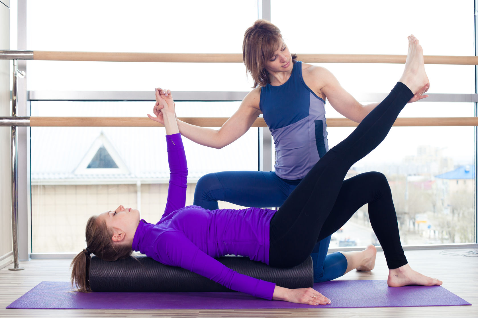
foam roller image

Return to Sport: Ankle Injuries

It’s coming up to sport season again. Footie boots, netball gear, orange slices and all of the aches and pains that are synonymous with kids sports. But how bad should an ache or a pain be before you go and see someone? Ankle sprains are the most common injury in active and sporting individuals. From a simple rolling of the ankle to a ligamentous tear, a physiotherapist can help. Mild sprain injuries result in a stretching of the ligaments which normally act as a passive restraint and support the ankle with twisting movements. As these ligaments stretch, it becomes easier and easier to re-injure the ankle. Ankle injuries can present with swelling, stiffness and pain.
Sometimes, pain may present in three ways; 1) pain only during activity, 2) pain upon rest following activity, or 3) pain upon waking the following morning after an activity. The presence of pain is the body’s way of warning you that a certain activity may be causing you harm, such as the stretching and tearing of the ligaments around the ankle. Pain following an activity indicates the presence of inflammation. Playing through pain may create a cycle of chronic inflammation which will hinder healing and prolong an injury. Inflammation is commonly treated with rest, ice, compression and elevation.
If your child is complaining of persisting pain and swelling around the ankle, it may be time to visit your local physiotherapist. With ligamentous stretching, the ankle becomes unstable and people may also experience a loss of balance. Commonly physiotherapists advise rest for 3-5 days in an acutely inflamed ankle, giving a compression bandage to help alleviate swelling. As pain diminishes with rest, your physio will prescribe exercises to help improve ankle stability to reduce recurrence risk.
The next big question is should your child use tape or a brace during sport? Ideally, the exercises prescribed by your physiotherapist will aim to improve stability. Though while the ankle is strengthening (which can take six to ten weeks) your child will be heading back to sport. There are pro’s and con’s to both options as seen in the table. Ultimately it is personal preference. If you are concerned about your children’s ankles in the upcoming sporting season, why not give our friendly physiotherapists a call at Hinteractive Physio on 5442 5556 and book an appointment.

PRO CON
TAPING
Not Bulky Skin irritation and pain with removal
Improves joint position awareness Expensive long term
Difficult to self apply
BRACING
Long lasting Bulky, difficult to fit in shoe
Easy to self apply Brace slipping with movement

Are Your Kids Finding Studying a Pain?

As we head towards the final part of the school year, our school kids are studying for school exams and carting ever increasing amounts of text books home. Unfortunately for a lot of kids, the long hours spent hunched over the books results in neck pain, back pain and headaches.  Sitting puts a lot of strain on the spine, especially when combined with the flexed postures often seen when using a computer or reading for prolonged periods.  Despite the need to study, there are ways to minimise the strain on the spine and prevent the pain and headaches from occurring.

Firstly, having a good posture and desk set-up for studying are essential.  Make sure that you are sitting with your bottom as far back in the chair as possible and if your lower back is sore, place a small cushion in the curve of your lower back to support it.  If your feet cannot touch the ground, get a small stool to place them on.  If you have an adjustable chair, ensure that the height of the chair allows you to look at your computer screen without looking up or down too far.  When reading textbooks, use a book support or place them on a cushion, bringing them closer to your eyes and requiring less forward bending of the neck and rounding of the shoulders.

Secondly, taking mini breaks every 30-60 minutes allows some relaxation and stretching of the muscles which become tight when studying.  Make sure you stand up, walk around and stretch the neck and back muscles.  These short breaks will allow you to continue to study for longer with less pain.  In addition to mini breaks, ensuring that you get regular exercise is important for numerous factors.  Exercise has many positive effects including keeping your muscles strong and flexible, improving your concentration and memory, as well as improving your sleep quality.  Despite these positive effects, many people stop exercising when they are studying due to time constraints.  As you can see though, a small time sacrifice for exercise will be outweighed by the benefits.

If you are suffering with neck or back pain from studying, get some assistance as soon as possible so that you are not sitting through exams in pain.  Seek assistance from your local qualified health professional. Hinteractive Physio has experienced therapists who can assess and treat the cause of your pain as well as offering a youth Yogalates class which combines yoga and pilates to improve posture, flexibility and strength. Hinteractive Physio can be contacted on 5442 5556 or visit our website: www.hinteractivephysio.com.au

Abdominal Muscle Separation – In pregnancy and Beyond

Abdominal Muscle separation (rectus diastasis) is a common occurrence during pregnancy, although many women are unaware that they have this condition.  Whilst this separation is a natural occurrence to accommodate a growing baby, it is important to prevent or minimize the degree of separation during pregnancy and monitor it carefully postnatally to ensure that it does not become problematic.

Diastasis Recti is simply a term for separation of the rectus abdominis muscle which is the outer “6 pack” muscle running from the breastbone to the pubic bone.  The muscle comprises a left and right half which is joined down the centre by thick connective tissue called the linea alba.  During pregnancy, this connective tissue stretches due to the growing baby and can become thinner and in some circumstances develop tears.  This causes the outer abdominal muscles to function ineffectively with the possibility of developing other issues such as lower back pain, core instability, incontinence, hernias, postural issues and the much complained about “mummy tummy”.

If you are pregnancy, there are some ways to assist in preventing a large diastasis.  Ensuring good posture, appropriate core muscle exercises, and avoiding excessive weight gain during pregnancy call all be beneficial.  Some people have a genetic predisposition to developing a large diastasis due to weaker connective tissue.  Pregnant women may notice a bulge or dome of the centre of the abdominal wall when they go to rise from a reclined position.  This is a tell-tale sign of an abdominal separation.

After having a baby, women should have their abdominal muscles assessed within the first month to determine degree and severity of any diastasis present.  Specific exercises can be prescribed by a physiotherapist along with supporting garments/braces to assist the recovery of the abdominal muscles.  In severe cases, surgery may be necessary but is generally not encouraged until you have finished having children unless hernias are present.  Early intervention offers the best chance of recovery so don’t delay in seeking help if you think that have may have an abdominal muscle separation.

Rebecca Steele is a physiotherapist with a special interest in the area of Women’s Health and can be contacted at Hinteractive Physio in Cooroy on 5442 5556.

Physiotherapy and Prostate Surgery

Prostate cancer is now the most diagnosed cancer in Australian men, and the second most common cancer to cause death in men, with over 20,000 men being diagnosed with the disease each year. Physiotherapists are encouraging people to speak more openly about men’s health and prostate cancer.  Local physiotherapist, Craig Steele wants to encourage men diagnosed with prostate cancer and who are to undergo surgery to seek pre and post surgery consultation with an appropriately trained physiotherapist.  Craig wants to raise awareness about the side effects of prostate cancer surgery, specifically incontinence and erectile dysfunction, as well as how a physiotherapist trained in pelvic floor muscle rehabilitation can help.  Up to 95% of men who have radical prostatectomy will experience incontinence, however most men will recover with the help of pelvic floor physiotherapy.


Whilst the awareness of treatment available for female incontinence is fairly well known, most men either don’t speak of their condition, or just assume that they need to put up with it.   Many men with prostate cancer believe that time will heal them post surgery and their continence will spontaneously return, however this is not always the case. With the help of a pelvic floor trained physiotherapist, men can regain their continence sooner.

.

According to the Australian Physiotherapy Association (APA), incontinence and erectile dysfunction are significant side effects arising from surgery to treat prostate cancer. Most men will be incontinent and have erectile dysfunction straight after the surgery and for around 5% of men, incontinence will persist a year later.


The APA encourages men to be proactive about “pre-habilitation” in preparation for prostate cancer surgery, using pelvic floor muscle exercises to help bladder control. Physiotherapy research has shown how pelvic floor exercises should be taught in order for men to benefit most from the program. These tailored exercises shorten the time and severity of men’s incontinence thus improving their quality of life.


If you or a loved one are about to undergo prostate surgery, or have already had an operation, physiotherapist Craig Steele from Hinteractive Physio can help you regain your pelvic floor muscle strength.  Craig has recently undergone specific training in this area with renowned physiotherapy leaders in the field.  He can be contacted on 5442 5556 to make an appointment.  As there is a great deal of education involved in this consultation, you may need to ask for an extended appointment slot.  Craig can also be contacted via email: hinteractivephysio@gmail.com

Vertigo? Physio can help!

Anyone who has suffered from vertigo will know that it can be extremely debilitating.  Often, sufferers don’t know that physiotherapy can help.  So what is vertigo…..

Vertigo refers to the sensation of movement, or spinning that occurs due to an issue with your vestibular system (balance system). Vertigo may be used to describe feelings of dizziness, lightheadedness, faintness, and unsteadiness. When your head moves, signals are transmitted to an apparatus in the inner ear called the Labyrinth that is made up of three semicircular canals surrounded by fluid. The labyrinth then communicates this information via the vestibular nerve to areas of the brain that control balance, posture and movement coordination.

The most common cause of dizziness is BPPV (Benign paroxysmal, positional vertigo). Others include: Inflammation in the inner ear, Meniere’s disease, neck joint dysfunction, vestibular migraine and acoustic neuroma. Rarely, vertigo can be a symptom of a more serious neurological problem such as a stroke or brain haemorrhage.

What is BPPV?

Benign paroxysmal positional vertigo (BPPV) is a condition characterised by episodes of sudden and severe vertigo (dizziness) associated with head movement. Common triggers include rolling over in bed, looking down and lifting your head to look up.

BPPV tends to come and go, with sufferers experiencing days or weeks of symptoms, then a period of no dizziness.

What Causes BPPV?

Inside your inner ear, semicircular canals set at different angles are filled with fluid. When your head is moved, the movement of the fluid inside these canals tells the brain exactly how far, how fast and in what direction your head is moving.

BPPV is caused by small crystals within the canals (calcium carbonate crystals).  These crystals are usually contained within a certain area of the inner ear, but sometimes become dislodged and disrupt the functioning of the balance system.  These crystals can become dislodged for various reasons including injury or degeneration, but in almost half of cases, the cause is unknown.

How can Physiotherapy Help?

Physiotherapists can perform an assessment to determine what side the problem is located on and can then perform a special technique to re-locate the problematic crystals.  A home exercise program is given as well to re-inforce the treatment.  Physiotherapists will also assess your neck to ensure that it is not contributing to dizziness.

It is important to see your GP if you are suffering from vertigo or contact a physiotherapist if you have been diagnosed with BPPV to reduce these symtpoms quickly.

Arthritis – don’t let the cold weather slow you down

The term arthritis means joint inflammation. The most common type of arthritis is osteoarthris (OA), which refers to degeneration of the joint surfaces. This degeneration leads to pain, stiffness, weakness, instability and sometimes locking and swelling. The most common joints to be affected are the large weight-bearing joints such as the knees and hips, as well as the spine, hands and feet. OA can be caused by an injury to the joint or from repetitive stress and strain. As the joint surfaces begin to degenerate, they become stiff, painful, inflamed and can sometimes begin to form bony growths called osteophytes. The muscles, tendons and ligaments surrounding the joints often become tight and tender and ultimately weak. As the cold weather approaches, sufferers of OA find that their symptoms often increase.

What can we do about OA?:

Well, once the damage is done to the joint, you can’t actually reverse that. But there are a great deal of treatment options to help manage the condition and prevent it from getting significantly worse.

  • Gentle exercise is important to keep the joints moving as freely as possible.
  • Strengthening the muscles around the joint also help to cushion the joint and prevent excessive negative joint movements. With arthritis of the spine, core stability exercises help to stabilise the damaged joints, much like braces and supports do.
  • Hands on physiotherapy techniques are very important for pain relief and optimising the muscle function around the joint. Physio’s use a variety of techniques such as joint mobilisation, muscle releases, acupuncture and electrotherapy to help minimise pain and improve function.
  • Hydrotherapy is another useful treatment technique which allows the joints to be exercised in a warm, relatively weightless environment.
  • Physiotherapists can also advise on appropriate braces or supports to help in more symptomatic cases. If you are suffering with the pain of arthritis, speak to a health professional about what can be done to help with your condition.

Rebecca Steele owns Hinteractive Physio and can be contacted on 5442 5556.

Avoiding Sporting Injuries – A Guide for Adults and Older Athletes

By Craig Steele (Sports Physiotherapist)

With winter well and truly upon us, we tend to start seeing the injuries flow into the clinic from various sports such as soccer, rugby, touch football and tennis. With Australians being encouraged to take up more activity and organised sport, it is timely to take a look at the causes of sports injuries and what we can do to minimise the risks of being injured in the first case.

The broad term of “sports injuries” refers to injury sustained whilst undertaking not only organised sport, but also physical activity/exercise. They can occur for a number of reasons including accidents, poor training surfaces, poor equipment, poor technique, lack of conditioning or inadequate warm up or stretching.

If starting a new sport, it is important to seek coaching to ensure that your technique is correct, you are using the correct equipment for your shape and size and you are exercising on the correct surfaces. Sports injuries can affect any part of your body, but typically relate to the musculoskeletal system (muscles, bones and joints). Some of the most common injuries are sprains, strains and fractures.

A sprain refers to stretching or tearing of the ligaments – the thick band that joins one bone to another. Sprains can range from Grade 1 (minimal stretch) to Grade 3 (complete tear of the ligament) and usually result from trauma such as a fall or knock from fellow competitors. It is important to know the Grade of sprain, as this dictates the course of rehabilitation. Symptoms include pain, swelling, laxity or inability to weight bear through the joint.

A strain is a pull or tear of the muscle or it’s tendon (attaches the muscle to the bone). It usually results from overstretching or overcontracting the muscle. Symptoms may include pain, muscle spasm and weakness.

How to Treat Sports Injuries:

If you experience a sporting injury, you should follow these principles until you can seek further assessment from a physiotherapist or suitably qualified health professional:
1. Rest – it is important to immobilise the area injured if the pain is severe, or reduce your activity as your pain dictates, especially in the first 24 hours.
2. Ice – apply ice packs to the injured area to reduce inflammation and swelling. Always put ice pack in a damp tea-towel to reduce the risk of ice burns and apply for no longer than 20min at a time. This can be applied regularly in the first 24-48 hours.
3. Compression – is used to support the area and reduce swelling. Bandages or tubular bandages such as “tubigrip” may be used
4. Elevation – elevating the injured area (especially for lower limb injuries) helps to reduce swelling in the area.

Tips for Preventing Injury

1. No weekend warriors! – don’t be inactive through the week, then go and play lots of physical sports or overdo the exercise. Try to spread your activity throughout the week.
2. Increase your exercise level gradually, especially when starting back at sport or exercise
3. Ensure you have a good technique – coaching may be necessary
4. Accept your bodies limits and modify your activities as needed
5. Use the correct safety gear
6. Maintain good overall fitness. It is important to combine cardiovascular activity with weight training and stretching exercises.
7. Seek treatment early on if experiencing pain or discomfort.

Remember, it is always easier to treat conditions early before they become chronic. Physiotherapists are trained to accurately assess sports injuries and implement an appropriate treatment program. Sports

Physiotherapists have undergone extra tertiary level training to refine their skills in assessing the biomechanics of sport, assessment of injuries and the implementation of thorough and sports related treatment programs.

Craig Steele has a Masters Degree in Sports Physiotherapy and owns Hinteractive Physio. He can be contacted on 5442 5556.

How to Stay Comfortable in the Saddle

Horse-riding is a very common activity among hinterland residents and is a great form of exercise. Back pain is also a common complaint, so, when is horse-riding beneficial and when it is harmful? What exercise should you do to help prevent back pain from riding and reduce your time off the horse.

Riding with a good technique uses most muscles in the body. Sitting on a saddle with a controlled, upright posture requires the core muscles to stabilise the body against the movements of the horse. This type of activity is great for the spine, as long as good posture and core stability are maintained. Problems occur when people are suffering with back pain and have lost their core control before they start riding, or when they are riding with poor posture and control or for longer periods than their fitness allows. Getting a horse to move in certain ways under command requires whole body strength along with core stability.

As physio’s we tend to see more injuries from looking after horses than we do from riding. The manual work required to look after a horse requires lots of lifting, bending and twisting. These repetitive actions are known to be harmful to the spine and often lead to injury. Be aware of your spine when you are cleaning and maintaining your horses. Use your legs more than your spine for lifting and turning and consider a back support if you have had a previous injury.

Most people ride on a weekend basis and may not be physically fit enough to cope with the length of ride and postural control required. Having good cardiovascular fitness is essential, so this type of exercise (eg. Walking, swimming, running, cycling) should be incorporated into your lifestyle. Core stability exercises such as pilates and fitball exercises will ensure that the postural muscles have the endurance required to support the spine during horse-riding. If you are already suffering with back pain, it is essential to have these muscles working well before you return to riding.

Seek advice from a physiotherapist early to avoid chronic back pain or time out of the saddle. Craig and Rebecca Steele operate Hinteractive Physio and can be contacted on 5442 5556.

Taking the Pain out of The Road Trip

Well school holidays are just around the corner and Christmas is less than a month away, so many of us will be packing up the cars and starting the traditional road trip. For many people though, the thought of long car drives makes them cringe, with the memories of back aches and neck stiffness flooding back quickly. But it needn’t be such a painful experience! With some easy tips and a few exercises, you’ll be arriving at your destination full of the Christmas spirit instead of a belly full of paracetamol.

But like most things, preparation is the key. If you have any niggling problems, get them sorted out before you go away. This way, you are starting your trip in good physical condition, rather than having to stop half way for urgent treatment. If you have room in your vehicle, be sure to pack your own pillows. This reduces those “wry” necks when you wake on the first morning of sleeping in a strange bed with a strange pillow. Next step- check your car set-up. Make sure that your seat is adjusted to provide optimum lumbar support and reduced arm straining to reach the steering wheel. Headrests need to be positioned so that they are not pushing the head forward, as this will cause neck pain and stiffness.

Most people, and especially those with back pain, will find that the in-built lumbar support in their car seats is not enough to support the lower back on a long drive. Lumbar rolls (round or d-shaped foam rolls) are a cheap and effective way to support the lower back while driving. They are simply placed in the curve of the lower back between the chair and the spine, and help to hold the natural lumbar curvature of the spine. Place your backside as far back in the chair as it will go, to help reduce “slouching”.

Try and stop every hour or so to reduce muscle and joint fatigue. Simply go for a short walk and try the following stretches.

  1. Place your hands in the hollow in the lower back and gently arch backwards as far as comfortable. Repeat this 5-10 times.
  2. Place one heel on the ground with the toes pulled back. Lean forward, poking the bottom out the back until you feel the stretch in the back of the thigh. Hold 20 seconds and sway sides.
  3. Stretch your neck by looking on a diagonal into your armpit. Hold 20 seconds and swap sides.

Obviously, if you have any specific problems you should consult a therapist before you go, to get more specific exercises for your body. So with regular breaks, not only will you be protecting yourself from driver fatigue, but you will be doing your body a favour.

For more information on back supports or exercises, contact your health professional. Craig and Rebecca Steele own Hinteractive Physio and can be contacted on 5442 5556.

Ladies – How’s your “core and pelvic floor?”

By Rebecca Steele

With Mother’s Day fast approaching, it is timely for all women to have a think about the health of their “core and floor”. The “core” muscles, refers to the deep muscles of the abdomen and lower back which have a stabilising role in the body and help to maintain a good posture. In addition to this important function, in women who are pregnant or have had a baby, they are vital for minimising the strain placed on the lower back while pregnant, and help to re-gain abdominal tone post pregnancy. A lot of women perform sit-ups diligently, but still have the tell-tale “pot belly” in the lower abdomen due to the lack of tone and endurance in the core muscles. The deep abdominal “core” muscles do not just return to normal function after they have been stretched, cut (caesarean section) or inhibited by moderate to severe back pain. They need specific exercises which can be taught to you by a physiotherapist or other suitably qualified fitness professional. The up-side to getting your core muscles working again is that the overall tone of your abdomen will improve, along with your posture.

The other essential health area for women to be aware of is their pelvic floor. Most women have heard of pelvic floor exercises but aren’t exactly sure of what they need to do and if they are doing it correctly. There are also some myths surrounding whether or not they should be performed while pregnant. The pelvic floor muscles are a sling of muscles which run from the tail bone at the back, through to the pubic bone at the front of the pelvis. The openings for the bladder, womb and bowel, all pass through the pelvic floor muscles, which are essential for the optimal functioning and support of the pelvic organs. Weak pelvic floor muscles can develop for numerous reasons, including pregnancy, childbirth, hormone changes, straining to pass bowel motions and chronic coughing. It is essential for all women, regardless of their age, to understand how to correctly perform a pelvic floor contraction, and ensure that they exercise the muscles regularly, especially when pregnant and following the birth. Like any muscle, if you don’t use it, you lose it. This is even more important as we age, as the hormone effects contribute to muscle weakness. Weak pelvic floor muscles can contribute to prolapses, incontinence and urgency symptoms in the bladder and bowel.

The “core” and “floor” (pelvic floor muscles) work together to provide a support network to the trunk from around and below. Squeezing up your pelvic floor while you are training your core muscles can assist the core muscles to work. However, if you suffer from pelvic floor weakness or incontinence, it is important to ensure you are performing an adequate pelvic floor contraction before doing core exercises, otherwise, more strain can be placed on the already weak pelvic floor. The good news is that pelvic floor weakness and incontinence can often be managed by a physiotherapist who has had extra training in assessing and treating conditions of the pelvic floor. Core muscle exercises can be provided by a physiotherapist, or suitably qualified health/fitness professional.

If you have any concerns about your “core and floor” please contact an appropriate health professional. Rebecca Steele co-owns Hinteractive Physio in Cooroy and can be contacted on 5442 5556.

Keep Your Body Merry This Festive Season

Well Christmas is nearly here, yet again. As physio’s, we often seen a great deal of people who either injure themselves over the festive season, or who find that their aches and pains increase over this period. So, why is that you ask? Well there are probably a number of reasons.

Firstly, we all tend to take a break from our regular exercise routines, and over indulge in some Christmas cheer. Regular exercise is essential for maintaining muscle strength, co-ordination and joint mobility. It takes a long time to improve muscle strength, but only a short period of rest to undo all of that good work. Without regular exercise, the receptors in our joints that send feedback to the brain don’t function as well as they should. This can increase the likelihood of an injury while sending some fast ones down the pitch in backyard cricket, or while hurtling down the ‘slip and slide’.

Alcohol is another major contributor to festive season injuries. Most people are aware of the balance effects of alcohol, so it is easy to see how it leads to increased falls and ligament damage. Ladies tottering around on high heels at Christmas parties are at a significantly higher risk of hurting an ankle or knee, especially if consuming alcohol. So ladies, no dancing in soaring high heels after a few champers’ at the Christmas party! At least take off the heels!

Overnight visits to relatives houses often involve sleeping in a strange bed, on a strange mattress and pillow. This often results in a wry neck upon waking, which is certainly not what you want during the Christmas break. Remember to take you own pillow when possible, and stretch your neck and back regularly to avoid muscle tightness.
So there you have it! Keep active, limit the alcohol (especially while wearing those killer heels) and stretch regularly to avoid a painful Christmas break. If you do happen to injure yourself this holiday period, seek help straight away. The sooner your injuries are attended to, the quicker you will be back to the holiday fun.

Craig and Rebecca Steele operate Hinteractive Physio and can be contacted on 5442 5556.

Achilles Heel of Football

By Craig Steele

With pre-season training well behind us, all football codes have strapped the boots on for the first couple of rounds and we are already seeing leg strains and injuries from both acute injuries and overuse injuries. Achilles tendon issues have been prevalent in these first few weeks of the season, as a result of training issues related to training surface and biomechanics along with training loads.

Most infamously we have recently seen the golden boot of David Beckham suffer an Achilles rupture, requiring surgical repair. His injury was not one of those acute sprains but rather an acute presentation of a long term degenerative condition of the Achilles tendon. The condition usually presents itself through a footballer’s career as episodic strains of the Achilles tendon known as “tendinosis”, and occasionally there is inflammation in the lubricating sheath of the tendon. The term “Achilles Tendinitis” is really a misnomer, as the tendon does not get inflamed itself but rather the surrounding structures. This repetitive micro-trauma of the tendon without adequate healing leads to the chronic condition of tendinosis, whereby the tendon suffers gradual degenerative breakdown. This is also true of all tendon structures of the body including the knee and shoulder.

Achilles tendon pain associated with an acute episode is due to inflammatory swelling around the tendon and poor tolerance of the tendon to further exercise loading. This is what we have been seeing in the early stage of the football season for all codes and is managed through appropriate treatment of causative factors and relative rest over the short term. However, it is the long term management that prevents progression of tendinosis into chronic, crippling states that may even progress to tendon tears or rupture. Physiotherapy is the mainstay of management for all tendon injuries that have not progressed so far along this injury continuum to require surgical intervention. Typical forms of treatment from a sports physiotherapist include massage, stretches, acupuncture and taping to deal with the acute presentation when there is swelling and inflammation in the surrounding tissues. But the keystone to good sports physio management is diagnosing the causative factors and assisting in future management of these issues when identified.

Experience with the different codes of football allows for greater understanding of the forces placed upon the lower limb during sport and the training requirements for coping with these strains on the Achilles tendon. Ultimately the recovery of the tendon relates to the strength and endurance of the whole leg as well as the core muscles of the hip. Flexibility is also an important issue for lower limb strength and stability during running and turning in football. For prevention or recovery from an Achilles tendon injury it is essential to address biomechanical issue arising from the foot and the dreaded footy boot which on average still lacks adequate support for the athletic foot. This often requires the correct fitting of footwear / boots and can even require intervention from orthotics inside the shoe to control poor foot posture and biomechanics.

If you have any concerns about your Achilles Heel or any other tendon injuries please contact an appropriate health professional. Craig Steele is a sports physiotherapist working at Hinteractive Physio in Cooroy and can be contacted on 5442 5556.

What is Physiotherapy and Can it Help Me?

As physiotherapists, we are often asked what we do and how it benefits the client. Physiotherapists primarily deal with movement. Another words, we ensure that your body functions and moves in an optimal, pain free and effective way. Problems with movement can be related to congenital (birth) issues, be a result of an accident, sporting or workplace injury, or be due to a major medical event such as a stroke or neurological condition. So whether you are young and active or elderly and sedentary, chances are at some stage in your life, you may benefit from physiotherapy.

Physiotherapists use a variety of techniques to ensure the optimal functioning of the muscles, joints and nerves. These include joint mobilisation or manipulation, muscle releases/massage, muscle retraining, exercise programs, electrotherapy to speed up healing and reduce inflammation, strapping and assistance with using various aids or braces. These techniques are used to help treat a variety of problems, including:

*Back pain
* Neck pain, headaches and whiplash,
*Arthritis
*Sports Injuries
* Rehabilitation from neurological disorders such as stroke, head injuries
* Upper limb pain including RSI, rotator cuff strains, tennis elbow etc
* Childrens problems such as cerebral palsy
* Pregnancy pain and weakness as well as incontinence

When you visit a physiotherapist, they will take a detailed history, perform a thorough physical examination, provide hands on treatment to address the problems found, advise you on the best course of management for your condition, and provide a home exercise program for you to optimise your physiotherapy consultation. The number of consultations required will depend on the severity and duration of your problems. If you have any queries about how physiotherapy may help you, please call your local physio who will be more than happy to answer any questions.

Rebecca Steele owns Hinteractive Physio in Cooroy and can be contacted on 5442 5556.